M. M. Tabbara1, L. Martinez1, J. C. Duque2, A. Paez1, G. Selman1, O. C. Velazquez1, L. H. Salman3, R. I. Vazquez-Padron1 1University Of Miami,Surgery,Miami, FL, USA 2University Of Miami,Medicine,Miami, FL, USA 3University Of Miami,Interventional Nephrology,Miami, FL, USA
Introduction: Intimal hyperplasia (IH) has been historically associated with improper venous remodeling and stenosis after arteriovenous fistula (AVF) creation. However, we recently demonstrated that IH does not explain brachiobasilic AVF maturation failure. Herein, we seek to evaluate whether IH plays a role in AVF focal stenosis. This study compares IH lesions in stenotic and nearby non-stenotic segments collected from the same AVF.
Methods: Focal areas of stenosis were detected in the operating room in patients (n=6) undergoing the second stage basilic vein transposition procedure. The entire vein was inspected and areas of stenosis were visually located with the aid of manual palpation and hemodynamic changes in the vein peripheral and central to the narrowing. Stenotic and non-stenotic segments were documented by photography before tissue collection (6 tissue pairs). Intimal area and thickness, intima-media thickness (IMT), and intima to media area ratio (I/M) were measured in hematoxylin and eosin stained cross-sections, followed by pairwise statistical comparisons.
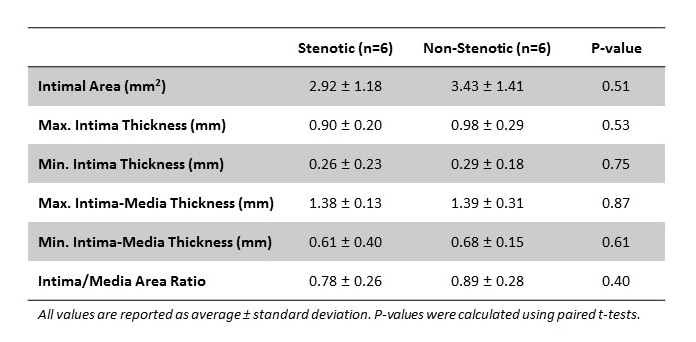
Results: The intimal area in stenotic and non-stenotic segments ranged from 1.25 to 4.82 mm2 and 1.67 to 5.30 mm2, respectively. There was no significant difference between these two groups (p=0.51). The maximal intima thickness (p=0.53), maximal IMT (p=0.87), and I/M (p=0.40) were also similar between both types of segments.
Conclusion: Despite the present limitations with the small number of samples, these findings concur with our previous study and suggest that IH is not associated with the development of focal venous stenosis in two-stage brachiobasilic fistulas. Our group is actively recruiting additional patients to extend these preliminary analyses.
