05.15 The Influence of Age on the Biology and Prognosis of Breast Atypia

A. R. Sergesketter1, S. M. Thomas2,3, A. M. Gupta1, O. M. Fayanju1,2, L. H. Rosenberger1,2, C. S. Menendez1,2, R. A. Greenup1,2, T. Hyslop2,3, E. S. Hwang1,2, J. K. Plichta1,2  1Duke University Medical Center,Department Of Surgery,Durham, NC, USA 2Duke Cancer Institute,Durham, NC, USA 3Duke University Medical Center,Department Of Biostatistics,Durham, NC, USA

Introduction:  Several prognostic variables and risk factors for invasive breast cancer have been shown to be age-related. However, the association between age and risk of cancer for women with high-risk breast lesions, such as atypia, is controversial. The aim of this study was to compare the prevalence of breast atypia and risk of upgrade or progression to breast cancer by age.

Methods:  Adult women diagnosed with breast atypia [lobular carcinoma in situ (LCIS), atypical ductal hyperplasia (ADH), or atypical lobular hyperplasia (ALH)] at a major academic institution from 2008-2017 were identified. Patients were stratified by age at initial atypia diagnosis: <50 years, 50-70 years, and >70 years. Differences between age groups were tested using Fisher’s Exact and Kruskal-Wallis tests. Logistic regression was used to estimate the association of age with risk of upgrade to or subsequent cancer diagnosis after adjustment.

Results: Among the 530 atypia patients (median age 54y) identified, 31.1% were <50y (N=165), 58.1% were 50-70y (N=308), and 10.8% were >70y (N=57). ADH was the most common finding in 75.1% of patients (N=398), followed by LCIS in 13.2% (N=70) and ALH in 11.7% (N=62). When comparing types of atypia by age, older women >70y were more likely to present with ADH, while women <70y were more likely to present with ALH or LCIS (overall p=0.04). Of the 471 women diagnosed with atypia by needle biopsy, 14.9% (N=70) were upgraded to breast cancer (invasive or in situ) at the time of surgical excision, which did not vary by age (p=0.33). Of the 70 women upgraded, 61.4% were to ductal carcinoma in situ (DCIS) and 37.1% to invasive disease, which was similar between age groups (p=0.4). The use of chemoprevention after an atypia diagnosis was also similar between all age groups (overall uptake 27.2%, p=0.55). During the follow-up of the remaining 458 women with atypia (median 49 months), 15.7% (N=72) developed a subsequent diagnosis of breast cancer (invasive and in situ) unrelated to the initial biopsy, which did not vary by age (p=0.66). Of those who subsequently developed breast cancer, 45.8% were diagnosed with DCIS and 54.2% with invasive disease, which was similar for all age groups (p=0.93). The unadjusted time to subsequent diagnosis of breast cancer did not vary by age (log rank p=0.41, Figure 1). After adjustment, age was not associated with upgrade to or subsequent diagnosis of breast cancer (both p>0.05).

Conclusion: Among women with breast atypia, age may influence the type of atypia diagnosed, but does not appear to be associated with risk of upgrade or subsequent progression to breast cancer.


05.13 Factors Associated With Neoadjuvant Therapy Use in Invasive Lobular Carcinoma of the Breast

R. A. Mukhtar1, J. M. Wong1, K. E. Fahrner-Scott1, C. Ewing1, M. D. Alvarado1, L. J. Esserman1, J. C. Boughey3, A. J. Chien4  1University Of California – San Francisco,General Surgery,San Francisco, CA, USA 3Mayo Clinic,General Surgery,Rochester, MN, USA 4University Of California – San Francisco,Hematology/Oncology,San Francisco, CA, USA

Introduction:  Although neoadjuvant therapy (NAT) increases breast conserving surgery rates in women with breast cancer, its effectiveness in invasive lobular carcinoma (ILC) has been questioned. Since surgeons must identify which patients may benefit from a neoadjuvant approach, we sought to determine factors associated with NAT in women with ILC. Additionally, we explored associations with neoadjuvant chemotherapy versus neoadjuvant endocrine therapy use. 

Methods:  We queried a prospectively maintained surgical database and identified 679 cases of clinical stage 1-3 ILC treated at our institution from 1981-2017. We collected patient characteristics, tumor size, subtype, stage, therapy, and era of treatment.  Data were analyzed in Stata 14.2 using t-tests for continuous variables, and chi-squared test for categorical variables. 

Results: NAT was used in 21.8% of cases, with 12.4% receiving neoadjuvant chemotherapy and 9.4% receiving neoadjuvant endocrine therapy.  Overall, women receiving NAT were significantly younger (57.3 vs 60.4 years, p = 0.0065), had larger tumors (2.9 vs 2.2 cm, p=0.0058), and had tumor subtype other than ER+ PR+ Her2- (p<0.001). NAT use significantly increased over time, initially consisting exclusively of neoadjuvant chemotherapy, but with an increasingly higher proportion of neoadjuvant endocrine therapy use in recent years (p=0.007, Figure).

We then analyzed ER+ Her2- cases (n=546, with 438 PR+ and 108 PR -), since NAT is questioned most in this group. Among these patients, NAT was significantly more common in younger women (56.8 vs 60.4 years, p=0.0033), and those with PR- disease (37.1% vs 17.8%, p<0.001). There was no difference in tumor size, treatment era, tumor grade, or histologic subtype. Lastly, within the ER+ Her2- cases who received NAT, those who received neoadjuvant chemotherapy were significantly younger (52.1 vs 61.9 years, p<0.0001), more likely to be premenopausal (45.1% vs 21.2%, p=0.01), and less likely to be diagnosed within the last 10 years (p=0.002).  Among premenopausal women with ER+ Her2- ILC receiving NAT, the only factor associated with receipt of neoadjuvant endocrine therapy versus chemotherapy was era of diagnosis, with significantly less chemotherapy in the last ten years (p=0.034).

Conclusion: Although many studies question the utility of NAT in ILC, our data show a striking change in management patterns over time, with a steady increase in NAT use and a shift from neoadjuvant chemotherapy to neoadjuvant endocrine therapy, even in premenopausal women. The long term impact of this new management strategy in ILC, and the utility of the information garnered about response to therapy, warrant additional study. 
 

05.12 Placement of Subcutaneous Central Venous Ports in Breast Cancer Patients: Does Side Matter?

C. A. Isom1, P. Bream3, R. N. Ahmed2, K. C. Gallagher2, S. Walia2, R. Kauffmann4  1Vanderbilt University Medical Center,General Surgery,Nashville, TN, USA 2Vanderbilt University,School Of Medicine,Nashville, TN, USA 3Vanderbilt University Medical Center,Radiology,Nashville, TN, USA 4Vanderbilt University Medical Center,Surgical Oncology & Endocrine Surgery,Nashville, TN, USA

Introduction:

The placement of subcutaneous central venous ports in breast cancer patients has become a common practice. Historically, ports have been placed on the side contralateral to the breast cancer due to concern about increased risk of complications with ipsilateral port placement. There have been only a few small studies evaluating complication rates between ports placed ipsilateral vs. contralateral to the breast cancer. We sought to determine if there was a difference in port complications or lymphedema rates by port location.

Methods:

A single institution retrospective review was conducted of adult (>18yrs) female patients undergoing central venous port placement for breast cancer treatment between 2012 and 2016. Patients that had ports placed by both surgery and interventional radiology were included. Patients were excluded if they had ports placed at another facility, their initial breast pathology was unavailable or treatment history was unavailable prior to their port placement.

Results:

A total of 581 females were identified with a mean age of 52.9 ±11.7 years. Ipsilateral ports were placed in 41 patients (7.1%). Ipsilateral ports were more likely to be placed via the internal jugular vein (56.1%) while contralateral ports were more likely to be placed in the subclavian vein (67.2%), p=0.002. There was no difference between type of breast surgery (p=0.997), axillary surgery (p=0.087) or administration of adjuvant radiation therapy (p=0.684) for patients that had ipsilateral vs contralateral ports. There was no difference in breast cancer stage at diagnosis but it did tend towards significance (p=0.0587). Ipsilateral ports were more likely to be on the right side, 73.2% vs 51.1% (p=0.006). Contralateral port were more likely to be placed for neoadjuvant therapy while ipsilateral ports were more likely to be placed for adjuvant therapy. Port complications requiring intervention occurred in 3(7.3%) patients with ipsilateral ports and 33(6.1%) patients with contralateral ports (p=0.73). Upper extremity lymphedema occurred in 8(20%) patients with ipsilateral ports and in 118(21.9%) of patients with contralateral ports (p=0.639).

Conclusion:

There was no difference in port complication or lymphedema rates between patients who had ports placed on the ipsilateral side compared to contralateral side for breast cancer treatment.


05.07 An Analysis of Active surveillance as a Treatment Modality in Ductal Carcinoma in Situ

A. C. Alapati1, T. A. James1  1Beth Israel Deaconess Medical Center,Surgery,Boston, MA, USA

Introduction: Ductal carcinoma in situ (DCIS) is a non-obligate precursor of invasive breast cancer. Current clinical trials are exploring active surveillance (AS) of DCIS. The purpose of this study is to characterize current practice trends in the use of AS.  The findings may inform clinical trials and provide insight into factors influencing adoption into practice.

Methods: The National Cancer Database was used to identify women diagnosed with DCIS from 2004-2015. Management with AS was defined as any patient not undergoing surgery, chemotherapy or radiation therapy. Multivariable logistic regression was used to assess patterns of AS.

Results: Of  84,281 women with DCIS, 342 (0.4%) underwent AS. Increased age, (OR 1.16), Hispanic/non-Hispanic black ethnicities (OR 1.90; 1.54), treatment at an academic facility (OR 1.76), non-private insurance (OR 1.29), lower grade (OR 1.34), and lower volume facilities (OR 1.60) were associated with higher use of AS. Patients with one or more comorbidities less frequently underwent AS compared with patients without morbidities. (OR 0.70) Residence distance from the treatment center showed no significance. Of all patients undergoing AS, 10.5% received hormonal therapy.

Conclusion: AS is currently an infrequently used treatment modality for patients with DCIS. We observed variations in AS based on age, ethnicity, facility type, facility volume, insurance status and tumor grade. The vast majority  of patients managed with AS did not receive hormone therapy. This information may further inform strategies for clinical trials, as well as quality of care in the management of DCIS.


05.08 Evolution of Indications for Nipple-sparing Mastectomy: Trends in Patient Selection Over A Decade

A. J. Bartholomew1, K. F. Griffith1, G. M. Lassiter1, S. Mehra1, M. Sosin2, D. L. Caragacianu1, S. C. Willey1, E. A. Tousimis1  2NYU Langone Health,Hansjörg Wyss Department Of Plastic Surgery,New York, NY, USA 1MedStar Georgetown University Hospital,Breast/Surgery,Washington, DC, USA

Introduction:  Rates of nipple-sparing mastectomy (NSM) continue to increase due to improved cosmesis and demonstrated oncologic safety. Successful outcomes have led to expanded patient selection in those who were traditionally considered to be non-ideal candidates. This study aims to characterize changes in patient selection for NSM over one decade at a single institution.


Methods: A single-institution, retrospective chart review identified all NSMs occurring from Jan 2008 through Dec 2017. Patient demographics traditionally included for patient selection criteria were collected including age, BMI, smoking history, breast size, ptosis, and history of radiation. Macromastia was defined as breast size larger than a C cup, obesity was defined as BMI > 30, smoking status was defined at the time of diagnosis, and ptosis was recorded as clinically significant if grade two or higher. Primary outcomes were differences in patient demographics evaluated by chi-squared and Student’s t-test across the decade in two bins (2008-2012 vs. 2013-2017). History of radiation was evaluated by breast, while all other variables were evaluated at the patient level.


Results: A total of 492 patients and 847 breasts were included in the final cohort spanning ten years. From 2008 to 2012, 157 (31.9%) patients underwent NSM, while 335 (68.1%) NSMs occurred from 2013 to 2017. The mean age of the cohort was 46.1 years (SD = 10.2), BMI was 23.6 kg/m2 (SC = 4.0), obesity was present in 39 (7.9%) of patients, 20 (4.1%) were current smokers, 92 (20.9%) had macromastia, and 51 (6.2%) breasts had a history of prior radiation. In the second half of the decade, patients were older (46.8 vs 44.7 years, p = 0.033), had larger BMIs (24.0 vs 22.8 kg/m2, p = 0.027), and a greater proportion had obesity (10.2% vs 3.2%, p = 0.004) and macromastia (23.8% vs 14.9%, p = 0.029; Table 1). There was no significant difference between the first and second halves of the decade in the proportion of active smokers (4.5% vs 3.9%, p = 0.758, respectively), patients with clinically-significant ptosis (33.3% vs 38.3%, p = 0.462), or breasts with a history of prior radiation (7.4% vs 5.7%, p = 0.389).


Conclusion: Over the last decade, indications for NSM have continued to expand to non-ideal patients. Our institution performed NSMs on a significantly increased proportion of women with larger breasts, higher BMIs, and older age throughout the last decade. 

05.06 Breast Density Does Not Differ Between Breast Cancer Receptor Subtypes

M. M. Goldbach1, D. I. Hoffman1, A. Malinovitch1, C. Huang1, M. Pomponio1, A. D. Williams1,3, S. M. Nazarian1, J. Tchou1,2  1Perelman School of Medicine at the University of Pennsylvania,Department Of Surgery,Philadelphia, PA, USA 2Perelman School of Medicine at the University of Pennsylvania,Abramson Cancer Center, Rena Rowan Breast Center,Philadelphia, PA, USA 3Lankenau Medical Center,Department Of Surgery,Wynnewood, PA, USA

Introduction:  Mammographic breast density (BD) is an established risk factor for breast cancer. The relationship between BD and breast cancer receptor subtype is unclear. We therefore aimed to evaluate differences in BD across tumor receptor subtypes at a large, academic medical center. Several studies have shown that triple negative breast cancers (TNBC) often present as interval breast cancer, i.e in between normal screening mammograms. We hypothesized that women with TNBC have denser breasts compared to women diagnosed with other breast cancer subtypes as classified by their hormone receptor (HR) and human epidermal growth factor receptor 2 (HER2) expression status.

Methods:  A retrospective database query from an institutional registry was performed to identify female patients diagnosed with primary breast cancer between 2009 – 2015 who also had a screening mammogram 6 ± 3 months prior to diagnosis at our institution. Patients were excluded if their breast cancers were noninvasive, metastatic at diagnosis, or had no receptor status information available. Patient demographic information, BD, and receptor status were collected. Categorical BD was measured using the Breast Imaging Reporting and Data System (BI-RADS) classification system (1 = almost entirely fatty, 2 = scattered fibroglandular densities, 3 = heterogeneously dense, 4 = extremely dense). A Chi-square test was performed to evaluate differences in BD across three tumor subtypes: HR+/HER2-, HER2+ (regardless of HR status), TNBC.  

Results: BD was assessed in 488 patients with invasive breast cancer. The cohort had a median age of 62 years (range 38 – 92). 58.2% of patients (284/488) were Caucasian, 37.9% (185/488) were Black, and 3.1% (15/488) were Asian. 78.5% of patients (383/488) had ER+/HER2- cancer, while 11.9% (58/488) had TNBC and 9.6% (47/488) had HER2+ cancer. The distributions of categorical BD between tumor receptor subtypes were summarized in Table 1. Overall, approximately 90% of patients (448/488) had a BI-RADS score of either 2 or 3. BD did not differ significantly across tumor receptor subtype (X2 2.88, p=0.823). 

Conclusion: In this pilot study, we report no significant difference in categorical breast density between breast cancer receptor subtypes. As categorical BD reporting is qualitative and has demonstrated inter- and intra-observer variability, we plan to quantify BD using our BD quantification software – Laboratory for Individualized Breast Radiodensity Assessment (LIBRA). We also plan to expand the cohort to increase our sample size. A multivariate logistic regression to control for other patient variables, including age, menopausal status, BMI, history of LCIS, and use of hormone replacement therapy is underway.


05.04 Cell Proliferation Genes but not Lymphatic Genes Associate with Breast Cancer Lymphovascular Invasion

M. Asaoka1,2, S. K. Patnaik1, F. Zhang1,3, T. Ishikawa2, K. Takabe1,2  1Roswell Park Cancer Institute,Surgical Oncology,Buffalo, NY, USA 2Tokyo Medical University,Department Of Breast Surgery And Oncology,Shinjuku, Tokyo, Japan 3Dartmouth Medical School,Lebanon, NH, USA

Introduction:

Lymphovascular invasion (LVI) is a significant prognostic factor in breast cancer. Multiple studies have reported that LVI is associated with increased number of tumor lymphatic vessels. However, the relationship of LVI with lymphatic-specific gene (LSG) expression is yet to be clarified. We studied which genes associate with LVI so that they may be useful as diagnostic markers.

Methods:

LVI status and mitotic scores of pre-treatment primary tumors were collated from pathology reports of 1046 breast cancer cases of The Cancer Genome Atlas (TCGA) project. Survival and other clinical data were obtained from TCGA publications. RNA sequencing counts from TCGA were normalized and log2-transformed to generate tumor gene expression data. LSGs, with ≥20x higher expression in lymphatic compared to blood endothelial cells, were identified from published studies. Clustering of cases by gene expression was examined with multi-dimensional scaling and tSNE methods. Top-scoring pair (TSP) and support vector machines (SVM) classifiers were used to quantify LVI-predictive value of LSG expression in leave-one-out cross-validation. Survival analysis was performed with Cox regression. Hallmark and Reactome sets were used for gene-set enrichment analysis. Categorical variables were compared by Fisher exact test, with P <0.05 deemed significant.

Results:

LVI was present in 242 (37.9%) of the 639 cases for which its status was noted. Patients with LVI demonstrated significantly worse outcome than those without for both 5-year progression-free (69.1% vs. 85.3%, P <0.01) and disease-specific survival (84.9% vs. 93.8%, P <0.01). Incidence of LVI was 2x higher in HER2 type compared to the other 3 subtypes (68.4% vs. 32.4%-37.8%; P = 0.04). LVI was also significantly related with late stage (I+II vs. III+IV: P <0.01; OR, 6.1), axillary lymph node metastasis (P <0.01; OR, 8.7), higher mitotic score (1+2 vs. 3: P <0.01; OR, 1.8), and higher nuclear score (1+2 vs. 3: P <0.01; OR, 2.1), but not with estrogen receptor status or tubular score (1+2 vs. 3). Of 84 LSGs, which included classical lymphatic markers like PROX1 and LYVE1, expression of only 3 was higher in LVI+ cases, and LVI+ and – cases did not cluster separately by expression of the 84 genes. LSGs also had poor predictive value for LVI in classification analyses (62% accuracy for both TSP and SVM). In global analysis, 950 genes were differentially expressed, with cell-cycle regulator gene SPDYC up-regulated the most in LVI+ tumors. Highest enrichments in the LVI+ group were observed for gene-sets related to cell proliferation.

Conclusions:

We did not observe any association of tumor lymphatic-specific gene expression with LVI. However, LVI was strongly correlated with expression of genes involved in cell proliferation. This was also reflected in association of LVI with higher mitotic scores, suggesting that highly proliferative cancer cells have a tendency to invade lymphatic vessels.

05.02 Outcomes for Patients with Residual Stage II/III Breast Cancer Following Neoadjuvant Chemotherapy

T. J. Stankowski-Drengler1, J. Schumacher1, B. Hanlon1, D. Livingston-Rosanoff1, K. Vande Walle1, C. C. Greenberg1, L. G. Wilke1, H. B. Neuman1  1University Of Wisconsin,General Surgery,Madison, WI, USA

Introduction:  Prior studies have demonstrated variations in rates of pathologic complete response (pCR) after neoadjuvant chemotherapy based on receptor status, with an association between pCR and survival for most receptor types. Fewer studies have examined survival and recurrence based on receptor status for women undergoing neoadjuvant chemotherapy who do not experience a pCR. Our objective was to evaluate differences in survival and distant recurrence for patients with residual stage II/III breast cancer following neoadjuvant chemotherapy by receptor type.

Methods:  A stage-stratified random sample of 11,360 patients with stage II-III breast cancer in 2006-2007 was selected from 1217 facilities in the National Cancer Database (NCDB) for a Commission on Cancer Special Study. We identified patients with residual pathologic stage II/III cancer after neoadjuvant chemotherapy. We excluded patients who did not receive standard of care therapy based on receptor status (i.e. endocrine therapy and/or Her2neu therapy). Medical record abstraction included distant recurrence as well as survival for 5 years post-diagnosis. Kaplan-Meier estimation was used to generate survival curves after neoadjuvant chemotherapy by receptor status.

Results: Our analytic cohort included 736 patients with residual disease after neoadjuvant chemotherapy. 58% of patients had ER or PR+/Her2neu- disease, 28% had ER and PR-/Her2neu- (triple negative) disease, and 14% had Her2neu+ (any ER/PR) disease. Median follow-up time was 7.2 years (0.6-9.4). Patients with triple negative cancer had the poorest 5-year overall survival (52% vs 82% Her2neu+ and ER or PR+/Her2neu-), and distant recurrence free survival (57% vs 72% Her2neu+ and 77% ER or PR+/Her2neu-) (Figure 1).

Conclusion: Patients with triple negative cancer who have residual disease after neoadjuvant chemotherapy have a significant risk of distant recurrence and mortality, when compared to patients with other breast cancer types. The relatively poor outcomes for patients with residual triple negative disease supports the consideration of additional adjuvant therapy as well as novel clinical trials for patients with triple negative with residual disease post-neoadjuvant chemotherapy. These data can be used by clinicians to counsel their patients regarding prognosis, specifically the relatively favorable prognosis for patients with options for targeted therapy in the face of residual disease.


04.20 Hypothermia Induces Lymphocyte Apoptosis and Dysfunction In Vitro

L. K. Winer1, A. M. Pugh1, V. Nomellini1,2, C. Caldwell1  1University Of Cincinnati,Division Of Research, Department Of Surgery,Cincinnati, OH, USA 2University Of Cincinnati,Division of Trauma, Critical Care, and Acute Care Surgery, Department Of Surgery,Cincinnati, OH, USA

Introduction:  Hypothermia can predict persistent lymphopenia in critically ill patients. In a murine model of intraabdominal sepsis that includes both surgical and antibiotic source control, these interventions were shown to normalize core body temperature, reverse lymphopenia, and improve survival of septic mice. However, the mechanisms linking hypothermia, lymphopenia, and mortality are still unknown. The objective of this study was to investigate the relationship between hypothermia and immunosuppression. We hypothesized that a hypothermic environment would induce lymphocyte apoptosis and dysfunction. 

Methods:  Single cell suspensions were prepared from the spleens of healthy CD-1 male mice. Lymphocytes were stimulated with an anti-CD3 antibody, and in vitro studies were performed at both 32 and 37° C. Lymphocytes were enumerated by flow cytometry. A fluorescent glucose analog was used to measure intracellular glucose uptake. Carboxyfluorescein succinimidyl ester (CFSE) was used to capture lymphocyte proliferation. Cytokines were quantified by a cytometric bead array assay. 

Results: To determine whether hypothermia directly leads to lymphopenia, Annexin V staining was performed. This showed an approximately 1.5 fold increase in splenic CD4 apoptosis in hypothermic (32° C) compared to normothermic (37° C) conditions (p<0.05). Because septic mice are susceptible to secondary infection, suggestive of immunosuppression, we next characterized whether the remaining lymphocyte population has altered function. We found that hypothermia is associated with a significant decrease in CD4 activation and cellular division based on CFSE quantification. To further delineate the mechanism underlying diminished CD4 division, we examined cell signaling and metabolic pathways. This revealed that hypothermic CD4 cells secrete significantly less IL-2, a cytokine imperative for lymphocyte proliferation. Correspondingly, there was a significant reduction in IL-2 receptor alpha chain CD25 expression on CD4 lymphocytes. Finally, splenic CD4 lymphocytes had a 25% decrease in glucose uptake under hypothermic conditions (p<0.05).

Conclusion: Our study demonstrates that hypothermia causes lymphopenia through a combination of increased splenic CD4 apoptosis and decreased cellular proliferation. The remaining pool of available splenic CD4 lymphocytes is dysfunctional, with significantly reduced activation and glucose utilization. These data suggest an integral relationship between hypothermia and immune compromise, stressing the importance of early source control and thermoregulation to reduce the morbidity and mortality from sepsis. 

04.19 Stop The Bleed (Stb): Development Of A Perfused Synthetic Cadaver Model

A. Gupta1, J. Rosenberg1, C. Villegas1, J. S. Curren1, R. Winchell1, M. Narayan1  1Weill Cornell Medical College,The Division Of Trauma, Burns, Critical And Acute Care Surgery,New York, NY, USA

Introduction:
As active shootings and other mass casualty incidents have become more prevalent, courses designed to teach basic hemorrhage control to laypersons have proliferated. In the current StB course, participants undergo hands-on training using a synthetic limb mannequin.  In a prior survey of 302 participants there was overwhelming sentiment that the mannequin was limited by its inability to demonstrate cessation of bleeding when hemorrhage control techniques were applied. We hypothesized that enhanced flow characteristics (pulsatile flow and flow at variable pressure) that can be stanched by StB techniques would improve the mannequin, and hence the experience and confidence of trainees.

Methods:
The mannequin was redesigned as a self-contained circulation model that could mimic both arterial and venous bleeding.  Different synthetic soft tissues were assessed for texture, thickness, compressibility, and durability.  Vessel material, construction, and placement were evaluated on their ability to mimic pulsatile blood flow and durability to repeated pressure, packing, and tourniquet applications.  Multiple mechanisms of simulating blood flow (gravity, pump) were also trialed. An 85 ml synthetic rubber capacity bulb with a 7.4 mm inner diameter tubing were used resulting in a stroke volume of 16 cc per hand stroke and pressure of 20-25 kPa or 150-187 mmHg. Finally, material cost was considered to facilitate low-cost, global distribution. The final mannequin resulted in an inexpensive, novel synthetic cadaver limb model that is equipped with vessels which mimic blood flow and provide a realistic wound on which to practice the hemostatic techniques of direct pressure, wound packing and tourniquet application taught in StB.

Results:
Nurse and physician educators conducted beta testing of the perfused mannequin. One-on-one interviews revealed positive feedback regarding both realism of the perfused mannequin and participants’ ability to obtain bleeding control using StB techniques. In addition, participants who trialed the mannequin reported an increased awareness of the rate of blood flow out of a wound, which in turn increased their sense of urgency in applying hemorrhage control techniques.

Conclusion:
Several training modalities are available to teach hemorrhage control techniques, varying from high-fidelity simulators, to animal models, to synthetic mannequins. In an effort to address shortcomings noted by participants in the current StB mannequin, we developed a novel perfused-bleeding mannequin that mimics both arterial and venous bleeding, responds appropriately to various hemorrhage cessation techniques, and is both inexpensive 
 

04.12 Coagulopathy: Putting Another “C” in PICS

L. K. Winer1, M. D. Goodman1,2, C. Caldwell1, V. Nomellini1,2  1University Of Cincinnati,Division Of Research, Department Of Surgery,Cincinnati, OH, USA 2University Of Cincinnati,Division Of Trauma, Critical Care, and Acute Care Surgery, Department Of Surgery,Cincinnati, OH, USA

Introduction: Patients who survive the acute phase of sepsis can progress to persistent inflammation, immunosuppression and catabolism syndrome (PICS), which is associated with organ failure and death. Although acute sepsis-associated coagulopathy is well described, coagulopathy of chronic critical illness is poorly defined. Given that acute sepsis leads to early depletion of coagulation factors, and PICS represents ongoing inflammation, we hypothesized that PICS is associated with consumptive coagulopathy driven by microthrombotic disease. 

Methods: Male CD-1 mice underwent 33% cecal ligation with a 25-gauge needle puncture and were sacrificed after 8 days, when mice concurrently display all PICS characteristics. Spleen megakaryocytes were enumerated using flow cytometry and hematoxylin and eosin (H&E) staining. Pulmonary microvascular thrombi were quantified with Martius Scarlet Blue (MSB) staining. Whole blood was anticoagulated, and complete blood counts and native and extrinsic thromboelastometry (NATEM, EXTEM) assays were conducted. Student t-tests were performed; p<0.05 was considered statistically significant.

Results: In our murine model of PICS, the spleen becomes markedly enlarged with increased splenocytes. To determine whether PICS splenomegaly was also related to megakaryocyte production, we used complementary methods of flow cytometry and histology. This demonstrated that during PICS, there is a greater than two-fold rise in splenic megakaryopoiesis (p<0.05) without a concomitant increase in peripheral platelets. Next, to determine whether the discrepancy between splenic megakaryocyte and platelet production was contributed to a consumptive process, we used microscopy. This revealed a nearly seven-fold increase in pulmonary microvascular thrombi in PICS compared with healthy mice (p<0.05). Finally, thromboelastometry was conducted to characterize coagulation. NATEM, a test of native coagulation, showed significantly delayed clot initiation time (CT), unchanged clot formation time (CFT), and significantly increased mean clot firmness (MCF) in PICS mice. When specifically examining the extrinsic pathway of coagulation (EXTEM), CT and CFT were decreased, and MCF was increased in PICS mice (all p<0.05).

Conclusion: Using a murine model of PICS, we demonstrated changes in splenic megakaryopoiesis, pulmonary microthrombi formation, and whole blood viscoelastic properties, suggesting a dysregulation of coagulation. Based on these findings, we propose that consumptive coagulopathy may constitute another cardinal feature of PICS. Altogether, these findings suggest that early identification of coagulation abnormalities may help predict and define PICS, as well as stratify those at greatest risk for subsequent organ failure and death.

02.10 Ex Vivo Assessment of Marginal Organs Using Dynamic Computed Tomography

M. K. Harris1, J. DiRito1,3, A. Feizi4, N. Boutagy2,5, A. Feher2,5, P. Yoo1, D. Haakinson1, D. Mulligan1, A. J. Sinusas2,5, G. Tietjen1  1Yale University School Of Medicine,Department Of Surgery,New Haven, CT, USA 2Yale University School Of Medicine,Department Of Internal Medicine, Section Of Cardiovascular Medicine,New Haven, CT, USA 3University of Cambridge,Department Of Surgery,Cambridge, CAMBRIDGESHIRE, United Kingdom 4Yale University School Of Medicine,New Haven, CT, USA 5Yale University School Of Medicine,Yale Translational Research Imaging Center,New Haven, CT, USA

Introduction:
Despite a high transplant waiting list mortality, many marginal organs are discarded based on incomplete and subjective data. Organ viability assessment relies on risk stratification of donor data and limited organ evaluation by visualization and frozen biopsy. Dynamic computed tomography (CT) is used in vivo to assess microvascular tissue perfusion and ischemia, factors related to damage sustained by transplanted organs. Dynamic CT has potential to provide objective, quantifiable data to improve marginal organ utilization. We adapted in vivo dynamic CT methods to an ex vivo setting to measure individual organ perfusion variability.

Methods:
CT was performed at the Yale Translational Research Imaging Center on a GE Medical Systems clinical CT scanner. 10 kidneys and 5 livers were obtained from a local pig slaughterhouse. Organs were dissected, flushed with cold Custodial HTK preservation solution, and stored on ice until imaging. DICOM images were reconstructed in Horos and analyzed using custom MATLAB code to measure rates of contrast enhancement and clearance throughout the organs over time.


Results:

Marginal organs are known to sustain microvascular changes. We expect that these microvascular obstructions impair flushing and promote tissue injury. We adapted dynamic CT for use with ex vivo organs by performing sequential scans during contrast administration with iohexol and during subsequent continuous flush. Using custom MATLAB code, we measured enhancement of two regions of cortex equidistant from the right and left sides of each organ and measured peak enhancement, rates of enhancement, and rates of clearance.

Figure 1A includes representative images of two kidneys. Region of interest (ROI) 1 and ROI 2 are measured in the cortex on the left and right sides of each organ and demonstrate varied contrast kinetics within organs. Kidney 3L had a slower rate of contrast accumulation, lower total enhancement, and faster rate of decay in the tissue (Figure 1B and 1C). These preliminary data may suggest that less microvasculature was engaged and that contrast primarily remained in the larger vessels.

Conclusion:
Dynamic CT of ex vivo organs can be used to directly quantify perfusion deficits and microvascular changes. Its use could be extended to assess marginal organs to more accurately identify viable organs and improve utilization. This technique also creates opportunities to potentiate other tools for assessing viability and to evaluate the effects of emerging techniques in ex vivo organ preservation, recovery, and treatment.


01.09 Optogenetic Stimulation of the Dorsal Motor Nucleus in the Brainstem Induces Increased Splenic Nerve Activity

A. M. Kressel1,3,4, T. Tsaava1, E. H. Chang1, Q. Chang1, V. A. Pavlov1,2, S. S. Chavan1,2, K. J. Tracey1,2  1The Feinstein Institute for Medical Research,Center For Biomedical Science,Manhasset, NEW YORK, USA 2The Feinstein Institute for Medical Research,Center For Bioelectronic Medicine,Manhasset, NEW YORK, USA 3Northwell Health,Department Of Surgery,Manhasset, NEW YORK, USA 4The Elmezzi Graduate School of Molecular Medicine,Manhasset, NEW YORK, USA

Introduction: The inflammatory reflex is a well-defined neural circuit composed of afferent and efferent vagus nerve fibers that both senses peripheral cytokine levels and regulates splenic tumor necrosis factor (TNF) production to maintain homeostasis. Previous studies have demonstrated that efferent vagus nerve signals originate both in the dorsal motor nucleus of the vagus (DMV) and in the nucleus ambiguus in the brainstem. The efferent arc of the inflammatory reflex relays functional signals from the efferent vagus nerve fibers to the splenic nerve, which, after entering the splenic parenchyma, modulates the release of acetylcholine from a subset of T-cells and a subsequent attenuation of TNF production from resident macrophages. Although this pathway has been extensively studied by us and other groups, the existence of a functional synapse between the vagus and splenic nerves remains controversial. Using optogenetics and neural recordings, we selectively studied the role of DMV cholinergic neurons in inducing splenic nerve activation.

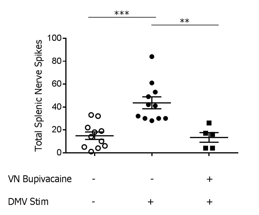
Methods:  Using stereotactic guidance, a fiber optic cannula was inserted into the DMV of transgenic mice expressing channelrhodopsin under the choline acetyltransferase promoter (ChAT-ChR2-EYFP) and stimulation was applied (473nm laser, 20Hz, 25% duty cycle, 3 minutes, n=12/group). Splenic nerve activity was simultaneously recorded using a cuffed two-channel electrode. Next, to determine the contribution of the vagus nerve in transmitting signals generated in the DMV to the splenic nerve, we measured splenic nerve activity before and after chemically blocking the vagus nerve. Following recording of splenic nerve activity during DMV stimulation, 0.05% bupivacaine was applied to the vagus nerve to prevent further signal transduction, and splenic nerve activity was recorded again during DMV stimulation.

Results: Splenic nerve activity was significantly increased over baseline (p=0.0002) during optogenetic stimulation of the cholinergic fibers in the DMV (Figure 1). Administration of bupivacaine on the vagus nerve significantly attenuated splenic nerve activity during optogenetic stimulation of cholinergic fibers in the DMV (p=0.0091), demonstrating a functional synapse between the vagus and splenic nerves.

Conclusion: These studies reveal that cholinergic fibers originating in the DMV induce splenic nerve activation via the vagus nerve. Knowledge of the neural circuitry of the inflammatory reflex, coupled with our previous work on endotoxemia, will allow for therapeutic interventions for patients with inflammatory conditions.

01.05 Glucose Increases Hexokinase-2 Expression and Glycolytic Capacity in Anaplastic Thyroid Cancer

A. Aggarwal1, Z. Yuan1, M. A. Nehs1  1Brigham And Women’s Hospital,Department Of Surgery,Boston, MA, USA

Introduction: Anaplastic thyroid cancer (ATC) is a fatal malignancy characterized by rapidly dividing tumor cells that demonstrate dependence on glycolysis for energy metabolism.  We therefore sought to investigate the role of the glycolytic enzyme Hexokinase II (HK2) which is over expressed in many malignancies, including ATC.  We hypothesized that a high glucose environment would promote HK2 gene expression and drive glycolytic energy capacity. 

Methods: We cultured ATC cell lines (JL30 and 8505C) in high (25mM) or low (3mM) glucose concentrations for 96 hours.  We analyzed HK2 expression by Fluorescent in-situ hybridization (FISH). We performed Seahorse XF Glycolytic stress tests to determine glycolytic reserve under each condition.  

Results: We found higher and more variable HK2 expression in both ATC cell lines in the high glucose medium.  JL30 had an average of 3.1 HK2 signals [Range 2-14] with high glucose environment versus 2.7 signals [Range 0-5] in the low glucose environment. Seahorse metabolic analysis revealed a glycolytic capacity of 245.8 mpH/min in the high glucose environment versus 55.9 mpH/min (p<0.001). 8505 had an average of 2.9 HK2 signals in the high glucose environment versus 2.7 in the low glucose environment. Seahorse metabolic analysis revealed a glycolytic capacity of 45.5 mpH/min for high glucose treatment versus 25.8 mpH in the low glucose treatment (p<0.01).

Conclusion: Here we demonstrate that a high glucose environment increases the expression of the glycolytic enzyme Hexokinase II and that this was associated with a higher metabolic glycolytic reserve.  These studies are suggestive that the metabolism of anaplastic thyroid cancer cells is influenced by glucose concentrations.  Future studies are needed to determine if hyperglycemia influences tumor biology in vivo.